Prophase I
n., [ˈprəʊˌfeɪz wŭn]
Definition: the first stage of meiosis I
Table of Contents
Organisms all use mitosis to create more cells in the body. Meiosis, a similar process, is used in some organisms to undergo the process of sexual reproduction. Within meiosis, there are many steps or phases, sometimes referred to as the meiosis stages. Prophase, Metaphase, Anaphase, and Telophase are the major stages of meiosis in which each phase occurs twice.
The stages of meiosis 1 (or meiosis I) are named after the first process and the same occurs for meiosis 2 (meiosis II). This includes prophase, which consists of prophase 1 (for meiosis 1) and prophase 2 (for meiosis 2). Let’s read on to find answers to what is prophase 1, what happens in prophase 1, and what is the importance of prophase 1.
Prophase 1 Definition
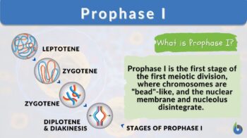
Prophase I is the first stage of meiosis I. This stage is characterized by five stages, namely leptotene, zygotene, pachytene, diplotene, and diakinesis, that make it up.
Prophase I highlights the exchange of DNA between paired chromosomes via a process called homologous recombination and the crossover at chiasmata (singular: chiasma) between non-sister chromatids. Thus, this stage is important to increase genetic variation.
This stage then ends with the disintegration of the nucleolus and the nuclear membrane. Prophase I precedes metaphase I, a stage that features homologous chromosomes aligning along a single plane in the center of the cell.
Before we move on and become acquainted with the various substages of prophase I, let us first understand some basic concepts.
Meiosis is a specialized form of cell division that ultimately gives rise to non-identical haploid gametes (or haploid sex cells). There are two successive nuclear divisions: meiosis I and meiosis II.
Each of them has four major phases. These are prophase, metaphase, anaphase, and telophase. Each of these phases is designated as I or II depending on where it occurs, i.e. in meiosis I or in meiosis II.
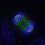
The paired homologous chromosomes in synapses are known as bivalents, and it is now clear that genetic recombination is what causes chiasmata to occur. These are visible under a microscope because of chromosomal condensation. One chromosome from each parent makes up the bivalent’s two chromosomes and four chromatids.
Watch this vid about prophase I:
Biology definition:
Prophase I is the first stage in the first meiotic division (meiosis I) characterized by having five sub-stages namely leptotene, zygotene, pachytene, diplotene, and diakinesis, and essential mainly for the exchange of DNA between homologous chromosomes via a process called homologous recombination and the crossover at chiasma(ta) between non-sister chromatids.
Variants: prophase 1; prophase one
See also: meiosis I, prophase
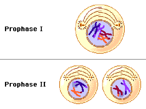
Meiotic Prophase I and Prophase II vs. Mitotic Prophase
Now, what is prophase? Is it the same as Prophase 1? How about Prophase 2? Prophase is that phase of mitosis and meiosis where the duplicated genetic material undergoes condensation in the nucleus.
In particular, the chromatin (a combination of DNA and proteins) of the parent cell, is condensed during prophase. (The condensed chromatins are referred to as chromosomes.) However, Prophase I differs from Prophase of mitosis and Prophase II of meiosis II in having these stages — leptotene, zygotene, pachytene, diplotene, and diakinesis.
These five stages of Prophase I make up the first stage of the first meiotic division. These distinct stages are, therefore, not observed in Prophase II and in Mitotic prophase. Nevertheless, all three are highlighted by the following features:
- Condensation of genetic material (chromatin), referred to as chromosomes
- Breaking down of nuclear envelope and nucleolus
- Microtubule-organizing centers (e.g., centrosomes) begin to move towards opposite ends of the cell, produce and organize the spindle
Conversely, Prophase I is unique in such a way that…
- Homologous chromosomes come together to form pairs and appear as bivalents
- Synaptonemal complex forms
- Crossing over occurs
Note it!
Prophase occurs in both meiosis and mitosis but Prophase 1 (as a stage) only occurs in meiosis.
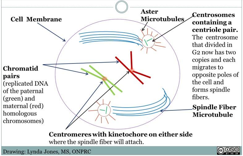
Centrosomes in Meiosis
For animal cells and animal-like cells where the centrioles in the centrosomes have a key role in cell division, the centrosome (a pair of centrioles) helps orient the chromosomes within the dividing cell so that by the end of mitosis or meiosis, both haploid daughter cells will have the appropriate number of chromosomes.
They do so by helping to form and organize spindle fibers that attach to the homologous chromosomes so as to line them up at the metaphase plate.
The centriole of the centromere replicates but it does so before prophase. New centriole assembles during the S phase of the cell cycle, which means during prophase I, the centrioles are already in pair and located near the nucleus. Nevertheless, the centrioles will disengage and be free to move to opposite poles (ends) of the dividing cell when the nuclear membrane disintegrates, which occurs during diakinesis.
Note that in humans, the centrioles (and centrosomes) are found in the spermatocytes but not found in many oocytes and ova. The spermatocytes (primary and secondary) are the male germ cells whereas the oocytes (primary and secondary) are the female germ cells that undergo meiosis (I and II) via spermatogenesis and oogenesis, respectively.
This means the primary spermatocyte would have two centrioles at the beginning of prophase I. Therefore, the two cells (secondary spermatocytes) formed after meiosis I will both have a single centriole, which will not be replicated again prior to prophase II. Thus, prophase I of the primary spermatocytes can be further characterized by having a centrosome with paired centrioles whereas prophase II of secondary spermatocytes will have a centrosome with only one centriole.
A prophase diagram can be viewed below in Figure 2.
Going back to human oocytes (together with some mammalian oocytes), the centrioles seem to have been lost when the oogonium grows into a primary oocyte at the stage of pre-ovulation. The oogonium still possesses intact pair of centrioles but when it grows into a primary oocyte that is bound for meiosis, particularly prophase I, the cell has seemingly lost the centrioles. And so, this leads later to spindle poles that are devoid of centrioles at metaphase I. This could mean there is another mechanism or pathway at play apart from the centriolar pathway in prophase I as spindle poles are still formed and organized at opposite ends during meiosis I.
The centriole in the sperm may not have directly contributed to the zygote following fertilization (which is also the time in which the oocyte completes meiosis and produce an ovum) but it could have provided the components for rebuilding centrioles, especially since the centrioles seem to rebuild and reappear at the spindle poles of the zygote (a diploid cell that serves as a starting cell of a multicellular organism) that divides via mitosis. (Gruss, 2018)

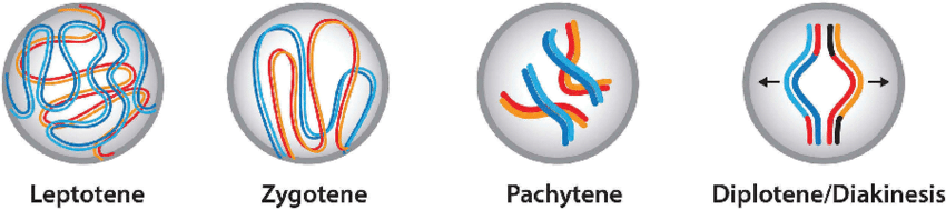
The Five Stages of Prophase I (Meiosis)
Prophase I is where the majority of the activities that distinguish meiosis from mitosis take place. When non-sister chromatids cross across, homologous chromosomes produce bivalents (or tetrads). In Prophase I, there are 5 separate substages:
- Stage 1: Leptotene – The chromosomes begin to condense and are connected to the nuclear membrane by their telomeres in leptotene.
- Stage 2: Zygotene – A synaptonemal complex forms between homologous chromosomes at the start of zygotene-synapsis.
- Stage 3: Pachytene – non-sister chromatids exchange genetic material (crossing over).
- Stage 4: Diplotene – Synaptonemal complexes vanish towards the conclusion of the diplotene synapsis, although homologous pairs remain linked at the chiasmata.
- Stage 5: Diakinesis – Chromosomes become fully condensed. The nuclear membrane and nucleolus break down in preparation for the next phase of meiosis I, which is metaphase I.
Several significant activities, including the pairing of homologous chromosomes and the reciprocal exchange of genetic material between homologous chromosomes, take place during these stages.
Prophase I meiotic progression occurs at rates according to sex and species. Until ovulation, several species halt meiosis in the diplotene of prophase I. In humans, prophase I arrest can last for decades until an egg swiftly enters meiosis I just before ovulation.
Stage 1: Leptotene
The chromosomes of prophase 1 under the microscope are visible. At higher magnification, they resemble “a string of beads” with nucleosomes acting as the beads. Some DNA may extend to about a centimeter in length, which is much too long for a cell nucleolus.
Therefore, it is packed utilizing unique proteins. Core histones are like the spools of sewing thread around which the DNA strands are wound. The nucleosome is a structure that is created when DNA has been wrapped twice around the core histone. This creates the appearance of a string of beads, with the unwound DNA serving as the string and the wound nucleosomes as the beads.
Each chromatid is incredibly near to the other, which frequently provides the impression that there is only one chromosome. It is also known that DNA double-strand breaks take place at the leptotene stage to prepare for recombination. In order to generate offspring with a wider range of alleles, recombination is the process by which the DNA of one chromatid is split apart and combined with that of another non-sister chromatid.
Crossing over during prophase 1 results in recombination. Given that the first stage is a fairly quick process in and of itself, leptotene is frequently referred to in conjunction with the subsequent stage as the leptotene-zygotene transition.
Stage 2: Zygotene
During meiosis, a chromosomal pair is created by joining a tetrad, or two homologous chromosomes made up of four chromatids. A synapsis is created so that two neurons can connect as a pair. The chromosomal pair is brought together and attached at a center point by filaments that resemble ladders.
The synaptonemal complex is made up of these filaments. The pair may only be referred to be a tetrad or bivalent once it has been joined. Once the synaptonemal complex has developed, crossing across can take place over it, however, in certain species, this complex is not necessary for recombination.
Stage 3: Pachytene
The process of crossing over and the ensuing recombination, in which a little amount of the genetic material from the parental DNA sequences is changed over to promote genetic variation, can proceed after a tetrad has formed.
The two chromatid strands that make up a single chromosome, known as sister chromatids, start to split from one another at this stage, however, the chromosomes still stay linked as a pair. They become considerably easier to distinguish under an electron microscope as a result.
The illustration below depicts the exchange of genetic material between two heterologous chromatids that are not sisters. The chiasma is the junction that connects two chromatids that are not sisters and permits the interchange of alleles. Only when the two sister chromatids are isolated from one another may chiasmata develop.
Stage 4: Diplotene
When the synaptonemal complex begins to deteriorate, as it does throughout the diplotene stage, the chromosomal pairs begin to split. They are unable to move too far away from one another since they are still linked by the chiasmata.
The unfinished meiosis I spindle machinery, which will be finalized during the prometaphase 1 that immediately precedes Prophase I, moves towards the opposing poles as a result of the two chromosomes’ repulsive characteristics.
Stage 5: Diakinesis
The chiasmata connections of diakinesis are located at the ends of the chromatid arms of the chromosome. Termination is the word for this entry. Chromosomes can no longer migrate toward the poles of the unfinished spindle construction because they are highly condensed and yet joined by chiasmata.
During meiosis I, more structural changes are made to prepare for the next phase. The nuclear envelope and nucleolus disintegrate. As a result, spindle remnants from mitotic cell division can travel together with centrioles (centrosome-forming microtubules), which are important for spindle formation. The primary components of spindle formation are microtubules found in the cytoplasm of the cell.
Prophase 1 arrest
All the oocytes required for future ovulations are present in newborn female mammals and birds, and these mammalian oocytes are halted at the prophase I stage of meiosis. Oocytes, for instance, are present at birth in humans because they develop within the fetus between three and four months after conception. The oocytes have four copies of the genome when they are in this prophase I arrested state, known as the dictyate, which may endure for decades. This is sometimes called meiotic arrest.
Prophase I arrest’s adaptive importance is yet not well understood. The halting of oocytes at the four genome copy stage, however, has been hypothesized to offer the informational redundancy required to repair damage to the germline’s DNA.
It indicates that homologous recombinational repair was utilized. Oocytes that are at prophase arrest have a high capacity for effective DNA damage repair. The capacity to repair DNA appears to be a crucial quality control mechanism in the female germ cell line and a vital factor in determining fertility.
Differences Between Animal and Plant Prophase
Plant cells lack centrioles, which is the primary distinction between prophase in plant cells and animal cells. Instead, foci at the polar opposites of the cell or chromosomes are involved in how the mitotic spindle apparatus is organized.
Preprophase, an extra stage in plant mitosis that culminates in the creation of the preprophase band, a structure made up of microtubules, is another obvious distinction. This band vanishes in plant mitotic prophase I.
Answer the quiz below to check what you have learned so far about prophase I.

Further Reading
References
- Genetics: From genes to genomes: Hartwell, Leland: Free Download, Borrow, and Streaming: Internet Archive. (n.d.). Retrieved July 23, 2022, from https://archive.org/details/genetics00lela_0/page/90/mode/2up
- Gruss, O. (2018). Animal Female Meiosis: The Challenges of Eliminating Centrosomes. Cells, 7(7), 73. https://doi.org/10.3390/cells7070073
- Meiosis prophase 1—The School of Biomedical Sciences Wiki. (n.d.). Retrieved July 23, 2022, from https://teaching.ncl.ac.uk/bms/wiki/index.php/Meiosis_prophase_1
- Meiosis Tutorial. (n.d.). Retrieved July 23, 2022, from http://www.biology.arizona.edu/cell_bio/tutorials/meiosis/page3.html
- Prophase | Learn Science at Scitable. (n.d.). Retrieved July 23, 2022, from https://www.nature.com/scitable/definition/prophase-189/
- Prophase—An overview | ScienceDirect Topics. (n.d.). Retrieved July 23, 2022, from https://www.sciencedirect.com/topics/agricultural-and-biological-sciences/prophase
- Stringer, J. M., Winship, A., Zerafa, N., Wakefield, M., & Hutt, K. (2020). Oocytes can efficiently repair DNA double-strand breaks to restore genetic integrity and protect offspring health. Proceedings of the National Academy of Sciences of the United States of America, 117(21), 11513–11522. https://doi.org/10.1073/pnas.2001124117
©BiologyOnline.com. Content provided and moderated by Biology Online Editors.