Spermatogenesis
n., plural: spermatogeneses
[ˌspɜːmətəʊˈdʒɛnɪsɪs]
Definition: The formation and maturation of spermatozoa
Table of Contents
Spermatogenesis Definition
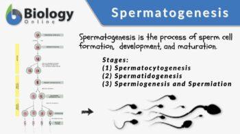
Spermatogenesis is the biological process of producing sperm cells. It occurs in the male gonad of a sexually reproducing organism. In this process, the undifferentiated male germ cells develop into spermatozoa through a series of events. It consists of these stages: (1) spermatocytogenesis, (2) spermatidogenesis, and (3) spermiogenesis and spermiation.
These stages occur in the seminiferous tubules of the testis. However, spermiation will continue in the epididymis where mature spermatozoa released into the lumen are propelled by the testicular fluid to the epididymis where they are stored (cauda region). In mammals, the spermatozoa will reach full motility (thus, maturity) when they reach the female reproductive tract. (Ref. 1)
In humans, spermatogenesis starts at puberty and continues throughout a lifetime. The entire process can take approximately 64 days. Spermatogenesis is the male counterpart of oogenesis in females. Spermatogenesis and oogenesis are the two forms of gametogenesis.
Etymology: The term spermatogenesis comes from the Greek sperma, meaning “seed” and from genesis, meaning “birth”, “origin”, “creation”.
Synonym: spermatogeny.
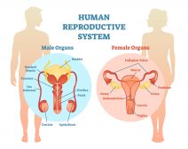
Male Anatomy and Germ cells
The testis (plural: testes) is the male gonad of animals, including humans. It is where mature sperm cells are produced and from where androgen hormones (mainly, testosterone) are released. In humans, it consists of testicular septa, testicular lobules, efferent ductules, rete testis, and seminiferous tubules.
The seminiferous tubules may be convoluted or straight. The convoluted seminiferous tubules are the twisted or curved tubules that serve as the site of spermatozoa production. The straight seminiferous tubules are the short straight tubules located between the convoluted seminiferous tubules and rete testis. Hence, they serve as a passageway for spermatozoa that come from the convoluted seminiferous tubules to the rete testis. The main cellular elements in the seminiferous tubules are the germ cells and the epithelial cell lining. (Ref. 2)
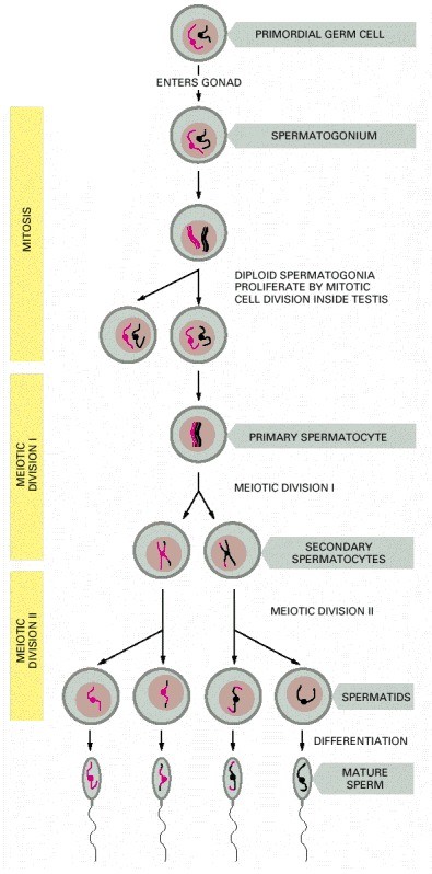
The germ cells are the cells that differentiate into sex cells (gametes). In spermatogenesis, the germ cells are spermatogonia (singular: spermatogonium), spermatocytes, and spermatids. Spermatogonia are the germ cells that divide mitotically to produce more spermatogonia. Some of them will differentiate into spermatocytes. Spermatocytes, in turn, are the germ cells that undergo meiosis. There are two types of spermatocytes: primary and secondary.
The primary spermatocyte is the germ cell that divides by first meiotic division and gives rise to two secondary spermatocytes. Each secondary spermatocyte enters the second meiotic division, giving rise to two spermatids. The spermatid enters spermiogenesis to become a spermatozoon (plural: spermatozoa).
See Table 1 for comparison.
Table 1: Male germ cells |
||||
|---|---|---|---|---|
| Spermatogonia | Primary spermatocytes | Secondary spermatocytes | Spermatid | Spermatozoon |
| The first germ cell stage; the rounded undifferentiated germ cells | The rounded germ cells that enter meiosis I | The rounded germ cells that enter meiosis II | The rounded germ cells that enter spermiogenesis | The streamlined, compacted sex cells with three distinct parts: head, mid piece, and tail (flagellum) |
| 2n | 2n | n | n | n |
| Location: Convoluted seminiferous tubules |
Location: Convoluted seminiferous tubules |
Location: Convoluted seminiferous tubules |
Location: Convoluted seminiferous tubules |
Location: Convoluted seminiferous tubules, then migrate to the epididymis |
| Stage: Spermato-cytogenesis |
Stage: Spermatid-ogenesis |
Stage: Spermatid-ogenesis |
Stage: Spermiogenesis |
Stage: Spermiogenesis and spermiation |
| Performs mitosis to give rise to other spermatogonia; Others develop into primary spermatocyte. | Performs meiosis I to give rise to secondary spermatocyte | Performs meiosis II to give rise to spermatids | Transform into non-motile spermatozoa | Differentiate into motile spermatozoa that may fertilize an ovum |
Apart from the germ cells, another major cell type found in the tubular lining is the Sertoli cell. (Ref. 3, 4) The Sertoli cells are epithelial cells of the seminiferous tubules. They are called nurse cells because they nourish the developing germ cells attached to them. Furthermore, they secrete testis-determining factor, a protein that concentrates testosterone in close proximity to the developing germ cells. They are also the cells that phagocytose residual cytoplasm of the maturing spermatid during spermiogenesis. In between the seminiferous tubules are Leydig cells. (Ref. 5) They are the gonadal cells that produce and secrete androgen hormones, including testosterone. The epididymis is the convoluted tubule that serves as the site of sperm differentiation and storage. (Ref. 6)
Hormonal regulation
The pituitary gland in the brain regulates the production of sperm cells and testosterone in the testes. In particular, the anterior pituitary releases luteinizing hormone (LH) that controls the release of testosterone. It is also the one releasing the follicle-stimulating hormone (FSH).
These pituitary hormones, together with the testosterone from the testes, regulate spermatogenesis. Testosterone is the primary male sex hormone and is responsible for the activation of the genes that are involved in spermatogenesis.
Stages
Spermatogenesis consists of stages: (1) spermatocytogenesis, (2) spermatidogenesis, and (3) spermiogenesis and spermiation. (Ref. 5) A spermatogenic cycle refers to the cycle that begins at one stage and ends with the same stage on a given segment of the seminiferous tubule at a certain duration. Different species have a different number of stages in a spermatogenic cycle.
Spermatocytogenesis
Spermatocytogenesis is the first stage of spermatogenesis. The spermatogonia in the basal lamina of the convoluted seminiferous tubule divide repeatedly by mitosis producing identical spermatogonia. Others move into the adluminal compartment of the convoluted seminiferous tubule and enter the first meiotic division as the primary spermatocyte. Both spermatogonia and primary spermatocytes are in diploid condition.
Spermatidogenesis
The next stage after spermatocytogenesis is spermatidogenesis. Thus, it is the intermediate stage of spermatogenesis. The highlight of this stage is meiosis, a type of cell division comprised of two succeeding divisions: first meiotic division (meiosis I) and second meiotic division (meiosis II).
In humans and other mammals, the seminiferous tubule is the only site in a male body where meiosis takes place. At this stage, the primary spermatocyte with its DNA duplicated enters meiosis I to give rise to two haploid secondary spermatocytes. Each of the secondary spermatocytes will immediately enter meiosis II to produce four genetically non-identical, haploid spermatids.
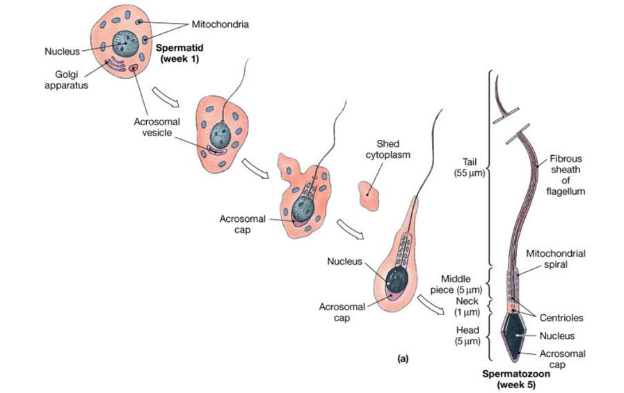
Spermiogenesis and spermiation
Spermiogenesis follows after spermatidogenesis. It is the stage of spermatogenesis wherein the spermatids become mature spermatozoa. The four phases of spermiogenesis are:
- Golgi phase
- Cap phase
- Tail phase
- Maturation phase
The spermatids will undergo events such as nuclear condensation, acrosome formation, mid-piece formation, and tail formation. However, the spermatozoa at this stage are not yet fully functional. Although they have acquired features essential in fertilizing an ovum (e.g. by becoming compacted and streamlined as opposed to the initial rounded shape with residual cytoplasm), they are not yet motile.
After spermiogenesis, the non-motile spermatozoa will leave the seminiferous tubules to travel to the rete testis in the mediastinum testis, to the efferent ducts, and finally to the epididymis. This migration of the spermatozoa to the epididymis is called spermiation. Since the spermatozoa are not yet motile at this stage, the Sertoli cells release testicular fluid to aid them as they travel.
The spermatozoa will gain motility in the epididymis. However, in mammals, full motility is achieved via capacitation, which takes place when the spermatozoa reach the female reproductive tract. (Ref. 1)
Events post-spermatogenesis
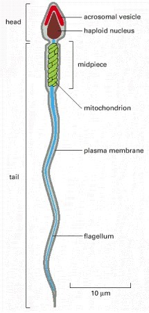
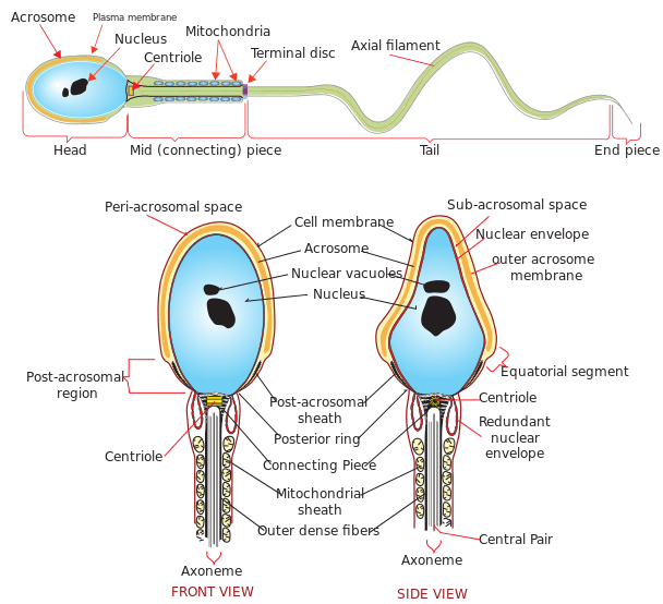
A motile and differentiated spermatozoon (sperm cell) has three distinct parts:
- the head
- the midpiece
- the tail
The head is capped with an acrosome. Inside the acrosome are enzymes that can break down the wall of the ovum at fertilization. The midpiece contains several mitochondria, i.e. the organelles responsible for the generation of ATP. This chemical energy is needed as the sperm cell prods its tail in search of the ovum. The tail is a flagellum containing an axoneme of 9 + 2 configuration.
The motile spermatozoa are stored in the epididymis until release (ejaculation). (Ref. 6) At ejaculation, the spermatozoa will move out of the epididymis, into the vas deferens, to the urethra, and out of the urethral orifice.
Table 2: Parts of spermatozoon |
|
|---|---|
 |
 |
| Credit: Alberts et al., (2002). NIH Books | |
Biological Importance
Spermatogenesis is a vital biological process. It is the means by which male gametes are produced. Through meiosis, it permits genetic recombination to increase genetic variations and thereby improve the gene pool. Any disturbance or interference in this process could lead to reduced fertility among males.
Some of the factors that could affect efficiency are temperature and nutrition. In humans, the normal scrotal temperature is approximately 34 °C, which is lower than the core body temperature (37 ° C). (Ref. 7) There should also be sufficient vitamins like vitamin B, E, and A. Spermatogenesis must be well regulated and kept within optimal conditions to make sure that the sperm cells will be devoid of lesions and abnormalities.
Answer the quiz below to check what you have learned so far about spermatogenesis.

Further Reading
References
- Visconti, P.E., Bailey, J.L., Moore, G.D., Pan, D., Olds-Clarke, P., and Kopf, G.S. (April 1995). “Capacitation of mouse spermatozoa. I. Correlation between the capacitation state and protein tyrosine phosphorylation” (PDF). Development. 121 (4): 1129–37. doi:10.1242/dev.121.4.1129.
- Male Reproductive 3 Seminiferous tubules. (2019). Retrieved from Okstate.edu website: https://instruction.cvhs.okstate.edu/Histology/mr/HiMRp3.htm
- Sertoli Cells. (2019). Retrieved from Yale.edu website: http://medcell.med.yale.edu/histology/male-reproductive-system-lab/sertoli-cells.php
- Griswold, M. D. (1998). The central role of Sertoli cells in spermatogenesis. Seminars in Cell & Developmental Biology, 9(4), 411–416. https://doi.org/10.1006/scdb.1998.0203
- Leydig Cells. (2019). Retrieved from Yale.edu website: http://medcell.med.yale.edu/histology/male-reproductive-system-lab/leydig-cells.php
- Male Reproductive System Lab. (2019). Retrieved September 17, 2019, from Yale.edu website: http://medcell.med.yale.edu/histology/male-reproductive-system-lab.php
- Ivell, R. (2007). Lifestyle impact and the biology of the human scrotum. Reproductive Biology and Endocrinology, 5(1). https://doi.org/10.1186/1477-7827-5-15
© Biology Online. Content provided and moderated by Biology Online Editors